
December 13, 2023 in Neural Networks
Revolutionizing Brain Tumor Detection: The CNN-Based Medical Imaging Breakthrough
SHARE: PRINT ARTICLE: https://doi.org/10.1287/orms.2023.04.03
A brain tumor is a typical cluster or proliferation of cells within the brain that can lead to disturbances in regular brain function and present diverse neurological symptoms. These tumors are categorized as either malignant or benign (as shown in Figure 1). Malignant tumors are cancerous, whereas benign tumors, though noncancerous, can still pose risks depending on their size and location. Currently, the medical community recognizes more than 120 different types of brain tumors [1]. Brain cancer is a severe medical condition that arises from malignant brain tumors that have the potential to spread and invade neighboring tissues through the spinal fluid. Their detrimental impact is often evident when they obstruct the normal flow of fluids within the human brain.

The American Cancer Society plays a crucial role in collecting, coordinating and examining cancer registries and mortality data obtained from the National Center for Health Statistics. According to their estimations for 2023, there will be approximately 1,958,310 new cases of cancer and 609,820 cancer-related deaths in the United States. On the bright side, it is predicted that there would be more than 19 million cancer survivors in the U.S. population by January 1, 2024 [2]. Among these, brain cancer is predicted to account for 24,810 new cases and 18,990 deaths [3].
This article presents a convolutional neural network (CNN)-based brain tumor detection model for classifying magnetic resonance imaging (MRI) images as containing tumors (YES) or not (NO). The deep learning model includes Conv2D, MaxPooling, Dense and Flatten layers (see Figure 2). Image segmentation is utilized to accurately detect cancerous patterns in MRI scans. Prompt early detection of tumors is vital to significantly improve patient outcomes. The model may achieve a 100% individual accuracy rate in quickly detecting small and large tumors. The implications of this model are significant, benefiting early diagnosis and treatment of brain tumors, leading to improved patient prognosis and preventing complications.

Medical Imaging
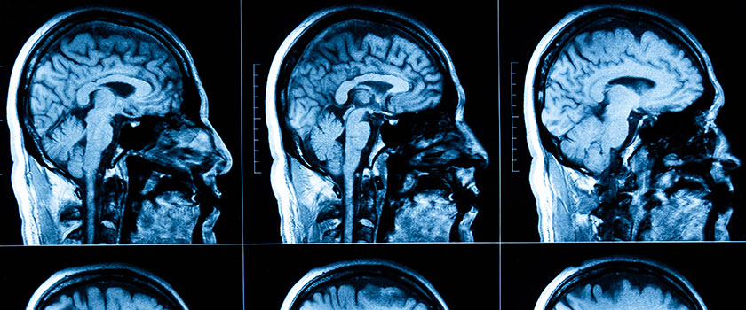
Medical imaging relies on sophisticated technologies to visualize internal structures within the body, greatly assisting in diagnosing, monitoring and treating various medical conditions. Several techniques, such as computed tomography (CT), positron emission tomography (PET), magnetoencephalography (MEG) and MRI scans, are utilized to aid in diagnoses. Among these techniques, MRI has gained widespread adoption because of its ability to differentiate between tissues and structures based on contrast levels. This noninvasive 3D imaging method effectively detects soft tissue or nonbony area anomalies. However, manual anomaly detection in MRI scans is labor intensive and prone to errors, which can seriously affect patients. To overcome these challenges, deep learning techniques have emerged as powerful tools for computer-based tumor detection using MRI scans. These techniques leverage the capability of an MRI machine to capture multiple images of the subject from different angles, contrasts and physical properties (as depicted in Figure 3). One deep learning algorithm, CNN, is discussed here and offers promising medical imaging and tumor detection advancements.

How Does an MRI Machine Work?
MRI utilizes powerful magnetic fields and radio frequency (RF) pulses to obtain images. It involves placing the body in a strong magnetic field, causing water molecules to align with the area when the magnets are on. Applying a strong RF pulse induces proton spin opposite the magnetic field’s direction. Once the RF is turned off, water returns to equilibrium, emitting RF signals that detectors capture and convert into images. The intensity of the emitted RF defines the tissue structure, which can be adjusted using scanner parameters. This enables the capture of multimodal images with an MRI scanner. Two crucial parameters in MRI imaging are as follows:
- TE time, also known as the time to echo, represents the duration between the RF pulse’s transmission and the echo signal’s reception during an MRI scan.
- TR time, the interval between consecutive pulse sequences applied to the same slice during the MRI process, is also known as repetition time [4].
Data Set
Data sets of brain tumor images comprise two categories: (1) YES and (2) NO. Here, YES means the MRI images of brain scans contain tumors, and NO means the MRI images of brain scans do not contain tumors. Both categories have 155 images of YES and NO. A balanced data set is crucial in a brain tumor detection model, ensuring sufficient examples of each class (tumor and nontumor) to effectively learn and generalize. When the data set is balanced, the model is not biased toward the majority class, which could lead to poorer performance in detecting the minority class (in this case, brain tumors). If the data set is imbalanced, in which one category (e.g., nontumor) significantly outweighs the other (e.g., tumor), the model may become biased and tend to classify most cases as nontumor, neglecting the tumor cases. As a result, the model’s accuracy and sensitivity in detecting brain tumors might be compromised. Data sets are like the brain of every model – the more accurate and proper they are, the better the model will be.
Methodology
A CNN-based brain tumor detection model was designed to assist medical professionals in categorizing tumors in brain scans. Using deep learning, the model aims to offer an accurate and efficient tool for tumor detection, reducing human error.
Convolution Neural Network (CNN)
Each layer used in the neural network for building this model is as follows (depicted in Figure 4).
- Data preprocessing: Before model building, medical images undergo preprocessing, including image resizing, normalization and data augmentation. Image resizing ensures uniform size, simplifying computational requirements and enabling efficient data processing. Normalization scales pixel values to a standardized range (typically 0 to 1), reducing the impact of intensity variations. Data augmentation expands the training data set using transformations such as rotations, flips and brightness adjustments, improving the model’s generalization and robustness to varying conditions.
- Conv2D layer: Conv2D is a fundamental building block of our CNN model. It applies filters to input images and generates feature maps by sliding over the image and highlighting specific patterns. These feature maps capture essential characteristics, enabling the network to learn more complex representations as the information flows through deeper layers. Conv2D plays a pivotal role in multiple computer vision tasks, including image classification, object detection and segmentation.
- MaxPooling2D layer: MaxPooling2D layers are essential in CNNs for down sampling feature maps, reducing complexity and controlling overfitting. These layers partition the maps into pooling windows and select the maximum value in each window. This down sampling decreases output size, making the model more efficient. MaxPooling2D helps the network focus on relevant features and generalize better to unseen data. Although it may cause some loss of spatial information, its benefits in computational efficiency and preventing overfitting make it a valuable tool in computer vision applications.
- Batch normalization: Batch normalization is a powerful technique in deep learning that enhances training by normalizing the activation functions of each layer. It mitigates the internal covariate shift problem, improving convergence and optimization. Batch normalization enables higher learning rates, leading to faster training and better performance. It acts as a regularizer, preventing overfitting, and reduces the need for manual hyperparameter tuning. This technique is a standard component in modern deep learning architectures, contributing to the success of training deep neural networks on complex tasks.
- Dropout layer: This layer is used to prevent overfitting in deep learning models. During training, it randomly deactivates neurons, promoting independent representations. This encourages the model to learn diverse features and improves generalization to new data. Dropout introduces regularization by reducing reliance on specific neurons and preventing coadaptation. It is a standard technique in training modern neural networks, enhancing their robustness and performance.
- Flatten layer: The flatten layer is crucial in deep learning models, converting multidimensional feature maps to a single vector, ensuring compatibility with dense layers. This step is essential because dense layers expect one-dimensional input. The flatten layer efficiently transforms the data for further processing in the fully connected layers, where global patterns and high-level decisions are learned.
- Dense (fully connected) layer: Fully connected layers interpret the flattened vector and generate the final output, determining tumor presence. They learn nonlinear relationships between features for accurate predictions. These layers are critical in medical image analysis, performing the last decision-making step in the model. The fully connected layers act as the decision-maker, converting the learned features into a conclusive prediction regarding tumor presence.

Performance Evaluation
We rigorously evaluated our CNN-based brain tumor detection model using a 90-10 training-testing split ratio on a diverse and representative data set. The model was trained with the AdaMax algorithmic optimizer and categorical cross-entropy loss function, achieving a remarkable training loss of 0.0018. This demonstrates the model’s efficient pattern learning and strong convergence during training. We assessed the model’s performance on a separate testing data set to ensure robustness, verifying its generalization capabilities. Combining a well-curated data set, efficient optimizer and appropriate loss function has resulted in a promising brain tumor detection model with potential applications in real-world medical scenarios.
Our CNN-based brain tumor detection model has shown great potential to assist medical professionals in accurately identifying brain tumors from medical imaging. Techniques such as data augmentation would be used for further work in improving this model. We aim to continue to develop a reliable and efficient brain tumor model that helps professionals ease their work and assist patients with their problems faster and more accurately. This model has a significant impact on the field of medical imaging analysis and improves patient outcomes.
In conclusion, the success of our CNN-based brain tumor detection model in accurately identifying brain tumors from MRI scans with a minimum accuracy rate of 99% per image is highly encouraging. By harnessing the power of deep learning techniques, we aim to contribute to the early detection and timely treatment of brain tumors, ultimately elevating patient outcomes and enhancing their quality of life. The model’s ability to learn intricate patterns from the data and its impressive convergence during training reinforces its reliability and effectiveness as a valuable tool to support medical professionals in medical imaging analysis.
As we refine this model, we are committed to further elevating its performance by incorporating advanced techniques such as data augmentation. Expanding the diversity of the training data through data augmentation will strengthen the model’s ability to generalize to unseen cases, thereby bolstering its real-world applicability. Our unwavering goal is to develop a dependable and efficient brain tumor detection model that can transform medical imaging. By facilitating faster and more accurate diagnoses, our model has the potential to significantly alleviate the burden on medical professionals, allowing them to focus more on patient care and treatment strategies. This technological advancement can pave the way for improved patient prognosis and more timely interventions, enhancing the overall quality of healthcare. With the promise of revolutionizing medical imaging, our CNN-based brain tumor detection model holds immense potential to make a substantial and positive difference in the lives of countless patients, underscoring the importance of continued research and innovation in the field.
References
- Kaur and A. K. Gill, 2017, “Review of Brain Tumor Detection Using Various Techniques,” https://doi.org/10.23956/ijarcsse/v7i4/0221.
- E. DeSantis, C. C. Lin, A. B. Mariotto, R. L. Siegel, K. D. Stein, J. L. Kramer, et al., 2014, “Cancer Treatment and Survivorship Statistics, 2014,” CA: A Cancer Journal for Clinicians, Vol. 64, No. 4, pp. 252-271.
- L. Siegel, K. D. Miller, N. S. Wagle and A. Jemal, 2023, “Cancer Statistics, 2023,” CA: A Cancer Journal for Clinicians, Vol. 73, No. 1, pp. 17-48, https://doi.org/10.3322/caac.21763.
- MRI, 2006, “Magnetic Resonance Imaging (MRI) of the Brain and Spine: Basics,” https://casemed.case.edu/clerkships/neurology/WebNeurorad/MRI Basics.htm.
Kushagra Agrawal is a computer science scholar in the School of Computer Engineering, KIIT Deemed to be University, Bhubaneswar, India. Connect with him on LinkedIn: https://www.linkedin.com/in/kushagra-agrawal-51a3121ab/. Nisharg Nargund is a computer science scholar in the School of Computer Engineering, KIIT Deemed to be University, Bhubaneswar, India. Connect with him on LinkedIn: https://www.linkedin.com/in/nisharg-nargund-6bb385255/.
([email protected])